The following are some diatoms I photographed using a DIC microscope and a digital camera connected to computer software:
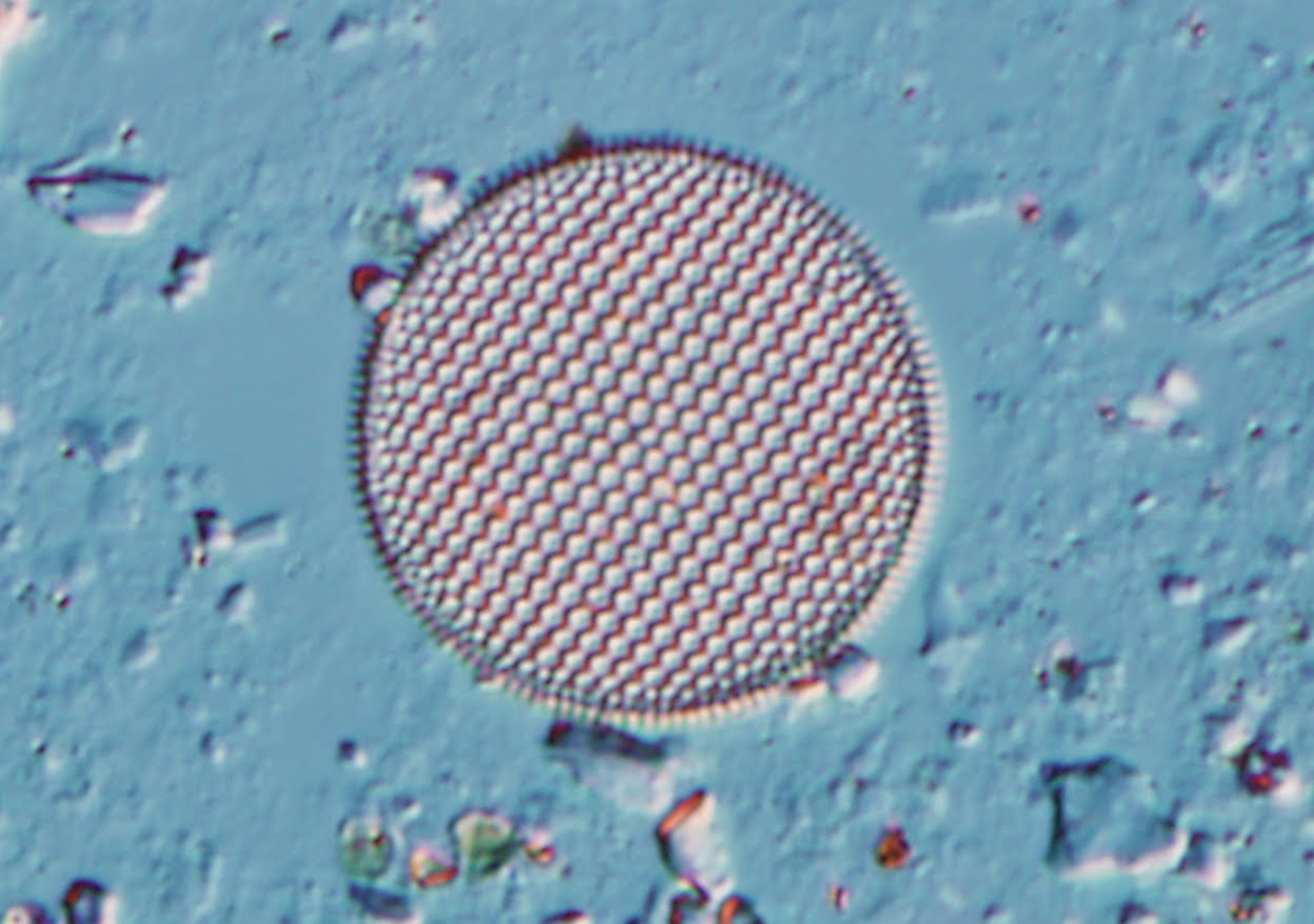


These were taken at 40X. Beautiful aren't they?? These were taken from prepared slides.
Today I went to some marine tanks on campus and learned to collect and prepare my own slides. I scraped a little sand off the bottom of a tank with some water in a vial and took it to a microscope.
I couldn't believe my eyes!! What appeared as ordinary salt water with a bit of sand was harboring a whole universe of swimming, twisting, moving, intricately shaped things! I saw tons of diatoms, as well as cyanobacteria, nematobes, and some microscopic shelled creatures.Beautiful!
Then I killed them.
I added a drop of Lugol's iodine soluiton and my universe stopped moving. The creatures were now fixed and preserved. I snapped some photos. Here's a lovely cyanobacteria. I took a picture using three different techniques: brightfield, darkfield, and DIC.
 |
| Brightfield (normal) microscopy |
 |
| Darkfield microscopy |
 |
| DIC microscopy |
Pretty cool, huh? Still learning to identify all the diatoms and microalgae. More on that next week!
Murderers!
ReplyDelete